

Walk into many operating rooms or a cath lab and you will often see a C-arm next to the table. The curved arm moves into place while the team checks where the hardware sits. It also helps guide an instrument as the case goes on. This is a key tool in C-arm radiology. It lets clinicians view images while the patient stays in the same position.
This guide explains what a C arm machine is. It also covers how it works. You can use it whether you are checking gear for a facility or looking at a C-arm for sale listing for purchase.
What Is a C Arm Machine?
An X ray C arm machine is a kind of mobile imaging unit. It gets its name from the C shape of its main frame. One side holds the X ray source, and the other side holds the detector. The frame stays fixed in that curve, so the parts keep pointing at the same area of the patient. This stays true while the arm turns, tilts, or shifts.
That setup is why C arm work is so useful during surgery. A technologist can swing the C to get an AP view, a lateral view, or an oblique view while the case is still going. The needed image can be taken within seconds. The patient does not have to be moved. The sterile field is also not disrupted.
How Does a C Arm X Ray Machine Work?
Every c arm x ray machine relies on a handful of core components working together in sequence:
- X-ray generator and tube – Produces the controlled X-ray beam
- Detector (flat panel or image intensifier) – Captures the X-rays that pass through the patient and converts them into a viewable image
- C-shaped gantry – Holds the tube and detector in alignment and rotates around the patient
- Mobile base and brakes – Supports transport and locks the unit in position during use
- Workstation and monitor – Processes, displays, and stores the resulting images
When the operator turns the system on, the generator sends a tight burst of radiation toward the patient. Hard parts, such as bone, along with metal implants, take in more of the energy. On the monitor, they look bright. Softer tissue comes back in grays.
The detector sits on the other side. It catches the radiation that gets through. Then it turns that signal into a digital picture right away.
C Arm Fluoroscopy vs. C Arm Radiography
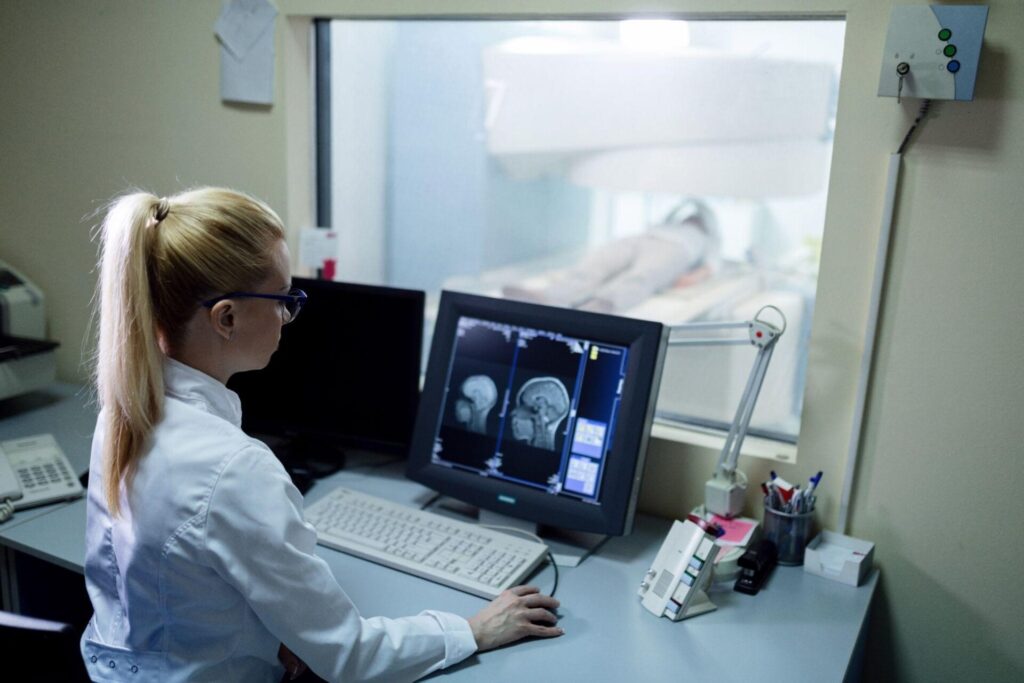
People mix up two related terms, so it helps to tell them apart.
C-arm fluoroscopy means continuous imaging in real time. You get a live video on a screen. The team can see the guidewire move, or see a break line line up as traction is applied, while it is happening. The unit sends out repeated low-dose pulses so the picture keeps updating.
C-arm radiography is different. It is one still picture. It is used when you want a clearer one-time view, like for records or for a close check. In that case, there is no need for ongoing video.
Most current C-arm machine systems can do both c arm fluoroscopy and c arm radiography. Clinicians can change the mode based on the step they are doing, using live guidance when they need it, and using a single exposure when they do not.
What Is C Arm Equipment Used For?
C-arm equipment supports a wide range of clinical applications, including:
- Orthopedic surgery – Confirming fracture alignment, fixation hardware, and implant position
- Vascular and interventional procedures – Guiding catheters, guidewires, and stents
- Urology – Assisting with access paths and instrument placement during procedures like PCNL
- Pain management – Supporting precise needle placement for injections and nerve blocks
- Emergency and trauma care – Providing imaging support when a patient can’t be moved to a fixed X-ray room
Because the imaging axis pivots as a single unit, surgical teams can check alignment from multiple angles within the same case, without adding significant time under anesthesia.
Features to Look for in Mobile C Arm Equipment
Not all c-arm equipment is built the same. When evaluating a system for an operating room, keep these factors in mind:
- Detector type – Flat-panel detectors (FPD) generally offer sharper image quality, wider dynamic range, and a more compact profile compared to older image intensifier (II) systems
- Detector size – Smaller detectors suit extremity and pain management procedures, while larger ones are better for spinal, abdominal, or vascular work
- Generator output and tube heat capacity – Determines how long the system can image before needing to cool, which matters most for longer procedures
- Maneuverability – Wheel design, braking system, and overall footprint affect how easily the unit moves through a busy OR
- Dose management features – Pulsed fluoroscopy, virtual collimation, and dose-area-product (DAP) monitoring all help minimize radiation exposure
- DICOM/PACS compatibility – Ensures smooth integration with existing hospital imaging systems
Getting these details right upfront makes a real difference in day-to-day workflow, not just on paper.
What to Consider When Evaluating a C-Arm for Sale
If you’re in the market for a c-arm for sale, a few extra considerations go a long way:
- Condition and service history – Ask for documented calibration and maintenance records
- Compatibility with your facility – Confirm the unit fits your OR layout, doorways, and table setup
- Regulatory compliance – Verify the system meets applicable radiation safety and type-approval requirements for your region
- Vendor support – Choose a supplier who offers installation, training, and ongoing service, not just delivery
A well-maintained system with a verified service history is often a smarter investment than a lower-priced unit with an unclear maintenance history, whatever c-arm for sale listing you’re comparing it against.
How Modern C Arm Systems Optimize Image Quality and Dose
Today’s C arm machine setup is based on one main idea. Make the picture as clear as you can, using the least radiation that still works. In most cases, that is done by:
- Pulsed fluoroscopy – Firing short bursts instead of a continuous beam, significantly reducing total dose
- Last-image-hold – Freezing the most recent frame so the surgeon can study it without additional exposure
- Virtual collimation – Adjusting the imaging field without extra radiation
- Flat-panel detectors – Offering better image clarity at comparatively lower doses than older intensifier-based systems
Combined, these features let clinicians get the visual guidance they need while keeping exposure as low as reasonably achievable, for both patients and staff.
Final Thoughts
A C arm machine system is more than one device. It is a full imaging setup. It includes the generator, the detector, and the screen. All parts work together to help surgeons see what they need during a case. If you are looking into c arm fluoroscopy, comparing detector options, or checking listings for a c-arm for sale, it helps to know what each part does. That knowledge supports better choices for both image quality and safe use.
Simons X-Ray is here to help. We are the trusted local source for diagnostic imaging gear in the Intermountain West. Over the years, we have spent more than 45 years selling and fixing X-ray equipment. That includes c-arm equipment used in surgery and in interventional settings. If you want to buy a C-arm, need help picking the right c arm x ray machine for your site, or want steady service after the sale, we can support you. We focus on practical help so your equipment stays safe and keeps working.
FAQs
What is a c arm machine, and how is it used in c arm radiology?
A C arm machine is a moving X-ray unit. It has a C-shaped frame that links the X-ray source to the detector. This setup helps staff take images during procedures. In c arm radiology, it can show images as the work happens. That means surgeons and interventional teams can avoid moving the patient between steps.
How does c arm fluoroscopy differ from standard c arm radiography during procedures?
C arm fluoroscopy enables continuous, live video imaging for real-time guidance, while c arm radiography takes a single still image for documentation or thorough investigation.
What features should you look for when choosing mobile c arm equipment for an operating room?
Key features are listed below. Detector type and size matter most for any c-arm equipment you consider. You also get the generator output. Maneuverability is another point to note. There are tools for dose control too. It also supports DICOM and PACS.
What are the top factors to consider when evaluating a c-arm for sale?
Check the state of the unit and the service history, compatibility with your facility, compliance with specifications, and the extent of vendor support received before committing to any c-arm for sale.
How does a modern c arm x ray machine optimize image quality while minimizing radiation dose?
Many c arm x ray machine systems run with pulsed fluoroscopy and use last-image-hold. They also apply virtual collimation. With flat-panel detectors, the picture stays clear. At the same time, the dose is kept low and as reasonable as possible.